Deeper inside, the use of chitooligosaccharides, in wound healing process. A computational approach Scientific paper
Article Sidebar
Main Article Content
Abstract
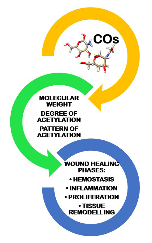
Chitooligosaccharides (COs) containing up to 10 monomeric units of N-acetyl d-glucosamine and/or d-glucosamine are water-soluble molecules revealing numerous biological activities and low toxicological profiles. Within this study, a computational approach has been used to predict the involvement of the COs having distinct chemical properties (molecular weight, deacetylation degree and acetylation pattern) in all the four wound healing phases: hemostasis, inflammation, proliferation and tissue remodeling. There are predictions, for the investigated COs, regarding their molecular targets and the biological activities that are reliant to the wound healing process. Furthermore, a molecular docking approach was used to assess the interactions of the investigated COs with the myeloid differentiation factor 2 (MD-2), a protein involved in the inflammatory processes. The investigation confirms the functional roles of the investigated COs in wound healing. The molecular targets predicted for the COs containing totally and partially acetylated units are galectins and selectins and those predicted for COs containing totally deacetylated units are fibroblast growing factors, the COs containing 3 units revealing the higher number of molecular targets. All these proteins are involved in mediating immune response, inducing cell division, growth and cell adhesion during the process of wound healing. All the COs containing from 2 to 8 monomeric units are able to interact with the MD-2 protein, the interactions being stronger for the COs containing 6 and 8 monomeric units. The interaction energies increase with the increasing molecular weight and with decreasing deacetylation degree and are reliant on acetylation patterns. Among the investigated COs, the totally acetylated COs containing 6 and 8 N-acetyl glucosamine units can be better inhibitors of the LPS binding to MD-2 protein. Consequently, mixtures of COs with distinct properties should be considered suitable candidates as adjuvants in developing scaffolds for the wound healing process.
Downloads
Metrics
Article Details

This work is licensed under a Creative Commons Attribution-NonCommercial-NoDerivatives 4.0 International License.

Authors retain copyright and grant the journal right of first publication with the work simultaneously licensed under a Creative Commons Attribution license 4.0 that allows others to share the work with an acknowledgement of the work's authorship and initial publication in this journal.


References
I. Aranaz, A. R. Alcántara, M. C. Civera, C. Arias, B. Elorza, A. Heras Caballero, N. Acosta, Polymers 13 (2021) 3256 (https://doi.org/10.3390/polym13193256)
B. Moghadas, A. Solouk, D. Sadeghi, Polym. Bull. 78 (2021) 4919 (https://doi.org/10.1007/s00289-020-03352-8)
L. A. M. van den Broek, C. G. Boeriu, C. V. Stevens, Chitin and Chitosan: Properties and Applications, Wiley, New York, 2020, pp. 232–238 (https://doi.org/10.1002/9781119450467)
X. Guo, T. Sun, R. Zhong, L. Ma, C. You, M. Tian, H. Li, C. Wang, Front. Pharmacol. 9 (2018) (https://doi.org/10.3389/fphar.2018.01412)
J. Li, D. Wang, S.-C. Chang, P.-H. Liang, V. Srivastava, S.-Y. Guu, J.-J. Shie, K.-H. Khoo, V. Bulone, Y. S. Y. Hsieh, J. Agric. Food. Chem. 69 (2021) 3371 (https://doi.org/10.1021/acs.jafc.0c06804)
D. L. Roman, M. Roman, C. Som, M. Schmutz, E. Hernandez, P. Wick, T. Casalini, G. Perale, V. Ostafe, A. Isvoran, Front. Bioeng. Biotechnol. 7 (2019) (https://doi.org/10.3389/fbioe.2019.00214)
D. L. Roman, V. Ostafe, A. Isvoran, Mar. Drugs 19 (2021) (https://doi.org/10.3390/md19030120)
D. L. Roman, M. Roman, H. Sletta, V. Ostafe, A. Isvoran, J. Mol. Graph. Model. 88 (2019) 41 (https://doi.org/10.1016/j.jmgm.2019.01.002)
D. L. Roman, V. Ostafe, A. Isvoran, J. Mol. Graph. Model. 100 (2020) 107676 (https://doi.org/10.1016/j.jmgm.2020.107676)
H. Jafari, K. V. Bernaerts, G. Dodi, A. Shavandi, Mater. Sci. Eng., C 117 (2020) 111266 (https://doi.org/10.1016/j.msec.2020.111266)
B. S. Park, J. O. Lee, Exp. Mol. Med. 45 (2013) e66 (https://doi.org/10.1038/emm.2013.97)
S. Viriyakosol, P. S. Tobias, R. L. Kitchens, T. N. Kirkland, J. Biol. Chem. 276 (2001) 38044 (https://doi.org/10.1074/jbc.M105228200)
Y. Qiao, Y. Ruan, C. Xiong, Q. Xu, P. Wei, P. Ma, X. Bai, Y. Du, Carbohyd. Polym. 82 (2010) 405 (https://doi.org/10.1016/j.carbpol.2010.04.079)
E. M. Jones, C. A. Cochrane, S. L. Percival, Adv. Wound. Care. 4 (2015) 431 (https://doi.org/10.1089/wound.2014.0538)
E. F. Pettersen, T. D. Goddard, C. C. Huang, G. S. Couch, D. M. Greenblatt, E. C. Meng, T. E. Ferrin, J. Comput. Chem. 25 (2004) 1605 (https://doi.org/10.1002/jcc.20084)
D. Gfeller, A. Grosdidier, M. Wirth, A. Daina, O. Michielin, V. Zoete, Nucleic Acids Res. 42 (2014) W32 (https://doi.org/10.1093/nar/gku293)
D. A. Filimonov, A. A. Lagunin, T. A. Gloriozova, A. V. Rudik, D. S. Druzhilovskii, P. V. Pogodin, V. V. Poroikov, Chem. Heterocycl. Cmpd. 50 (2014) 444 (https://doi.org/10.1007/s10593-014-1496-1)
H. M. Kim, B. S. Park, J. I. Kim, S. E. Kim, J. Lee, S. C. Oh, P. Enkhbayar, N. Matsushima, H. Lee, O. J. Yoo, J. O. Lee, Cell 130 (2007) 906 (https://doi.org/10.1016/j.cell.2007.08.002)
A. Grosdidier, V. Zoete, O. Michielin, Nucleic Acids Res. 39 (2011) W270 (https://doi.org/10.1093/nar/gkr366)
S. Salentin, S. Schreiber, V. J. Haupt, M. F. Adasme, M. Schroeder, Nucleic Acids Res. 43 (2015) W443 (https://doi.org/10.1093/nar/gkv315)
N. Panjwani, Ann. Transl. Med. 2 (2014) 89 (https://doi.org/10.3978/j.issn.2305-5839.2014.09.09)
M. Subramaniam, S. Saffaripour, L. Van De Water, P. S. Frenette, T. N. Mayadas, R. O. Hynes, D. D. Wagner, Am. J. Pathol. 150 (1997) 1701 (https://pubmed.ncbi.nlm.nih.gov/9137094/)
H. Tomita, Y. Iwata, F. Ogawa, K. Komura, K. Shimizu, A. Yoshizaki, T. Hara, E. Muroi, K. Yanaba, S. Bae, M. Takenaka, M. Hasegawa, M. Fujimoto, S. Sato, J. Invest. Dermatol. 129 (2009) 2059 (https://doi.org/10.1038/jid.2008.446)
D. S. Allen-Gipson, J. Wong, J. R. Spurzem, J. H. Sisson, T. A. Wyatt, Am. J. Physiol. Lung. Cell. Mol. Physiol. 290 (2006) L849 (https://doi.org/10.1152/ajplung.00373.2005)
A. Ala, A. P. Dhillon, H. J. Hodgson, Int. J. Exp. Pathol. 84 (2003) 1 (https://doi.org/10.1046/j.1365-2613.2003.00235.x)
R. H. Quarles, J. Neurochem. 100 (2007) 1431 (https://doi.org/10.1111/j.1471-4159.2006.04319.x)
S. Yamakawa, K. Hayashida, Burns Trauma 7 (2019) 10 (https://doi.org/10.1186/s41038-019-0148-1)
W. Li, Y. Li, S. Guan, J. Fan, C.-F. Cheng, A. M. Bright, C. Chinn, M. Chen, D. T. Woodley, EMBO J. 26 (2007) 1221 (https://doi.org/10.1038/sj.emboj.7601579)
J. Guo, C. Chang, W. Li, Expert. Rev. Proteomics 14 (2017) 665 (https://doi.org/10.1080/14789450.2017.1355244)
S. Gingis-Velitski, A. Zetser, M. Y. Flugelman, I. Vlodavsky, N. Ilan, J. Biol. Chem. 279 (2004) 23536 (https://doi.org/10.1074/jbc.M400554200)
M. D. Bagood, R. R. Isseroff, Int. J. Mol. Sci. 22 (2021) (https://doi.org/10.3390/ijms22116135)
L. Thomas, S. Mathew, S. Johnson, Inform. Med. Unlocked 20 (2020) 100406 (https://doi.org/10.1016/j.imu.2020.100406)
G. R. Vasta, Nat. Rev. Microbiol. 7 (2009) 424 (https://doi.org/10.1038/nrmicro2146)
K. Masuoka, M. Ishihara, T. Asazuma, H. Hattori, T. Matsui, B. Takase, Y. Kanatani, M. Fujita, Y. Saito, H. Yura, K. Fujikawa, K. Nemoto, Biomaterials 26 (2005) 3277 (https://doi.org/10.1016/j.biomaterials.2004.07.061)
H. Quan, F. Zhu, X. Han, Z. Xu, Y. Zhao, Z. Miao, Med. Hypotheses 73 (2009) 205 (https://doi.org/10.1016/j.mehy.2009.02.018)
M. Jiang, X. Zhuge, Y. Yang, X. Gu, F. Ding, Neurosci. Lett. 454 (2009) 239 (https://doi.org/10.1016/j.neulet.2009.03.042)
M. Jiang, Q. Cheng, W. Su, C. Wang, Y. Yang, Z. Cao, F. Ding, Neurochem. Res. 39 (2014) 2047 (https://doi.org/10.1007/s11064-014-1387-y)
V. Voinchet, P. Vasseur, J. Kern, Am. J. Clin. Dermatol. 7 (2006) 353 (https://doi.org/10.2165/00128071-200607060-00003)
T. Liu, L. Zhang, D. Joo, S.-C. Sun, Signal. Transduct. Target. 2 (2017) 17023 (https://doi.org/10.1038/sigtrans.2017.23)
H. Huang, Y. Zou, H. Chi, Drug. Des. Devel. Ther. 12 (2017) 67 (https://doi.org/10.2147/DDDT.S148064).